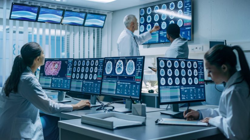
Orthopedic Imaging
Imaging tools and techniques are constantly evolving. They are used to diagnose and treat a wide range of orthopedic conditions and are helpful in better understanding an injury or ailment. Orthopedic surgeons, orthopedists, and sports medicine specialists always have imaging solutions at their disposal to accurately diagnose injuries or disorders affecting the muscles, bones, tendons, ligaments, spine, or cartilage. These tests are then used to help create an effective treatment for patients.
Types of Orthopedic Imaging

By creating images of certain parts of the body with the help of orthopedic imaging, our specialists can:
- Diagnose the causes of symptoms more accurately
- Use the images as a guide during surgical procedures or treatments
- Monitor the patient’s health following an appropriate treatment
- Assess and closely follow the progression of chronic diseases and the healing of injuries
The best resource for orthopedic imaging is Orthopedic Imaging: A Practical Approach, a book trusted by both radiologists and orthopedic surgeons for authoritative, comprehensive guidance on the interpretation of musculoskeletal images, having nearly 4,000 high-quality conventional radiography, ultrasound, CT, 3D CT, dual-energy CT, MRI, PET, PET/CT, PET/MRI, and other imaging techniques. The following tells you more about orthopedic imaging, including MRI, X-rays, CT scans, and functional imaging.
X-Rays
X-rays are the most common type of test used by an orthopedic surgeon. They are created by shining a small amount of radiation through a body part, and then the image is gathered on an X-ray film and processed. Orthopedists use X-rays to diagnose fractures, joint dislocations or to make further investigations to determine whether you could suffer from other conditions (such as arthritis or osteonecrosis). Most orthopedic practices now use digital X-rays over traditional film X-rays, as they use much less radiation. Moreover, the resulting images are more significant, precise, and color-enhanced, providing a more accurate diagnosis.
Computed Tomography (CT) Scan
A Computed Tomography (CT) scan is very much like an X-ray, offering a 3D view of a part of the body. While they provide very detailed pictures of the anatomy of joints or bones, they are known to be time-consuming. The patients are also subjected to more radiation than an X-ray. CT scans are used by doctors to diagnose a bone or spinal tumor and can also detect trauma to the spinal cord, pelvis, abdomen, or chest.
MRI (Magnetic Resonance Imaging)
MRI is a crucial component of orthopedic diagnostic services and uses a magnetic field to provide detailed images of bony structure and soft tissue. Orthopedists use MRI to diagnose a torn muscle, cartilage, ligament, herniated disc, osteoarthritis, hip or pelvic problems, tumor-like infections, and metabolic and endocrine disorders. The magnetic field makes the molecules in the body vibrate, translating them to detailed, 3D pictures that show what’s inside a tendon, ligament, or cervical spine. In patients with Alzheimer’s disease, MRI scans of the entire brain can accurately assess the rate of hippocampal atrophy. There is no radiation exposure during an MRI procedure, but it can be claustrophobic for some patients. There are different types of MRI scanners, from open MRI, closed MRI, whole-body scanners to extremity-specific scanners (only for legs, hands, arms).
Musculoskeletal Imaging & Ultrasound
Ultrasound imaging is a valuable technique in orthopedics. The procedure is entirely safe for all patients, including children, pregnant women, and people who have metal implants or tattoos. The ultrasound technology uses sound waves to produce pictures of joints, ligaments, tendons, muscles, or soft tissue. The musculoskeletal images are perfect when wanting to diagnose the cause of real-time tendon and ligament pain. At times, the ultrasound may detect tiny tears that the MRI might miss.
Both musculoskeletal imaging and ultrasound imaging are used to help diagnose numerous soft tissue injuries, including the following:
- Sprains and strains
- Tendon, ligament, or muscle tears
- Benign and malignant soft tissue masses (lumps/bumps) and tumors
- Ganglion cysts
- Hernias
- Nerve entrapments such as carpal tunnel syndrome
- Foreign bodies in soft tissues such as splinters or glass
DEXA Bone Density Scan
Dual-energy X-ray absorptiometry (DEXA) is the most common test used for measuring bone density and diagnosing osteoporosis. The narrow X-ray beams help doctors diagnose infection, cancer, or arthritis. Other imaging modalities are arthrography or bone scans. While arthrography uses a fluoroscope and contrast iodine solution to help diagnose the cause of unexplained joint pain, a bone scan can detect any areas of inflammation in a bone or body part. Doctors use orthopedic imaging, such as image-guided injections, to provide more accurate treatment to their patients. During these treatments, the doctors use ultrasound to visualize better the exact area where treatment should occur.
Our Doctors’ Approach
At Ceda Orthopedic Group, we are a group of highly skilled radiologists who have exceptional skills in orthopedic imaging. Our team works intensely to diagnose your condition and to find the proper treatment. Additionally, we use the most advanced orthopedic imaging technology available today, including conventional radiography, CT, or MRI. We treat a full range of conditions, from standard and complex cases to more acute and chronic ones. Whether you have muscle, bone, or joint pain, we will offer the best services to help you recover and enjoy your life.
We provide thorough medical documentation, utilizing detailed examinations and diagnostic services, including computerized tomography, magnetic resonance imaging, videofluoroscopy, and nerve conduction studies.
For the convenience of our patients, we offer transportation to and from the center. Our goal is to get you back to full health and your normal routine as soon as possible. We will make sure our orthopedic services will meet all your needs and will help you recover. To schedule an office visit or imaging appointment, call us at (305)646-9644 without hesitation! It’s time to get back on your feet and enjoy life as it was meant to be!
